SEAVER LAB IMAGES
All Photo Credit: Elaine Seaver Lab
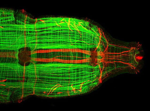
Capitella teleta
Confocal image of posterior regeneration in a juvenile worm. Capitella can regenerate complex tissues including nervous system, musculature, digestive system, and epidermis. Muscle fibers are visualized with filamentous action staining (green) and the nervous system is visualized with an anti-acetylated tubulin antibody (red). Ventral view.
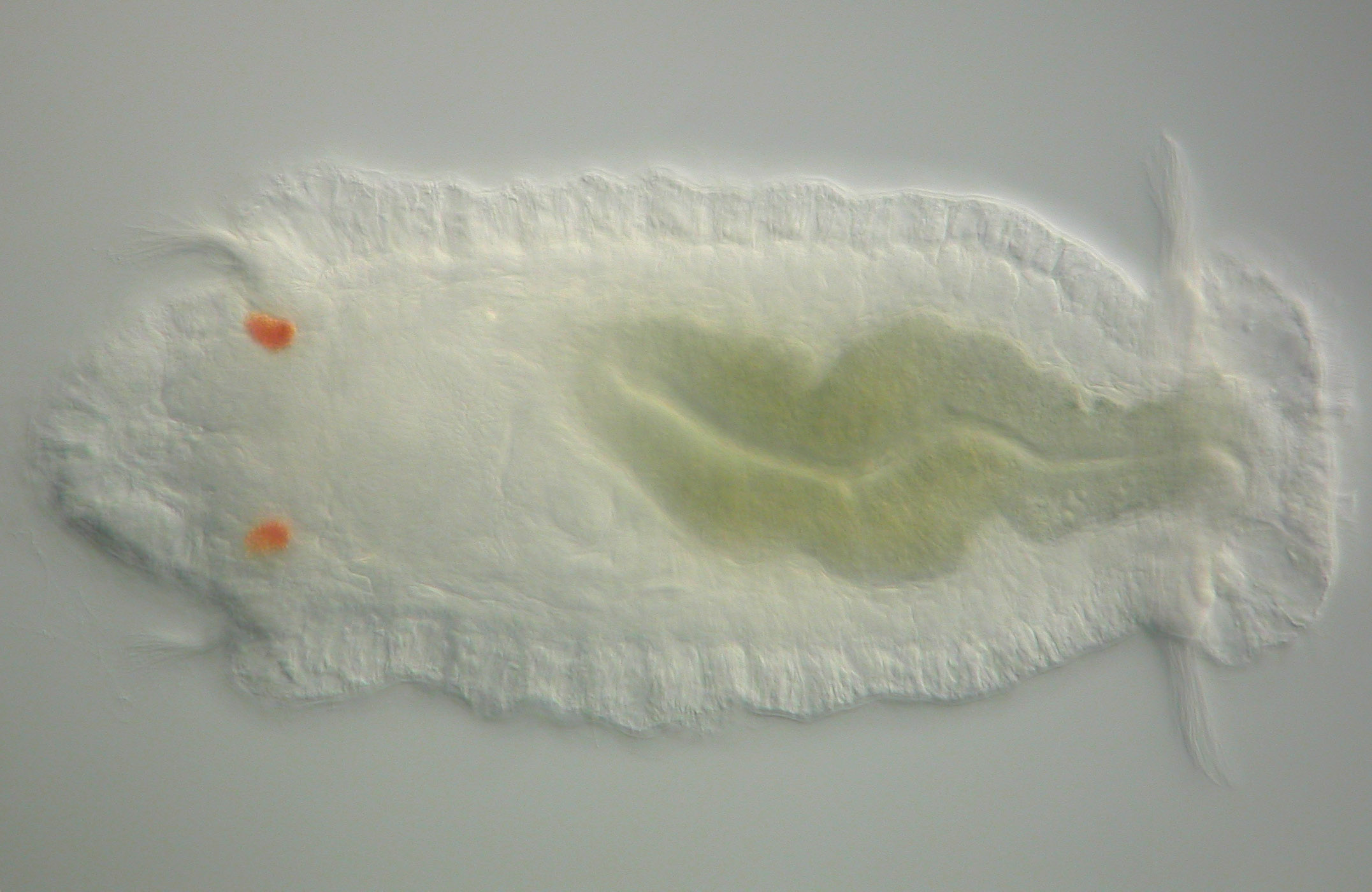
Capitella teleta
Early stage larva. The larval eyes are visible by a dark orange pigment. The larva swims by ciliary beating of two bands of cilia that extend around the circumference of the animal.
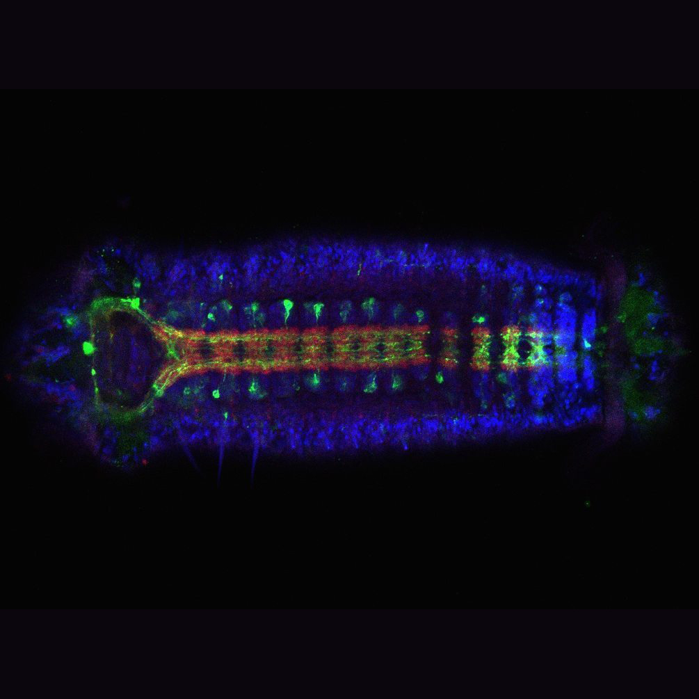
Capitella teleta
Centralized nervous system of a larva (ventral view). The nervous system is visualized by labeling with an anti-FMRF antibody (green) and an anti-synapsin antibody. Nuclei are in blue.
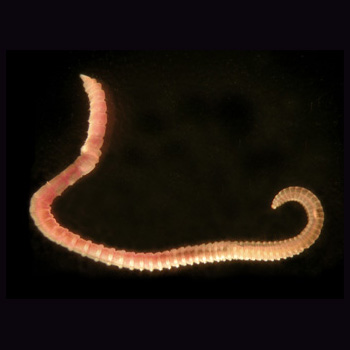
Capitella teleta
Adult worm showing the segmented body that is typical of annelids. The head end is on the left side of the image. The blood cells in the coelomic cavity give the animal a pink/red color.
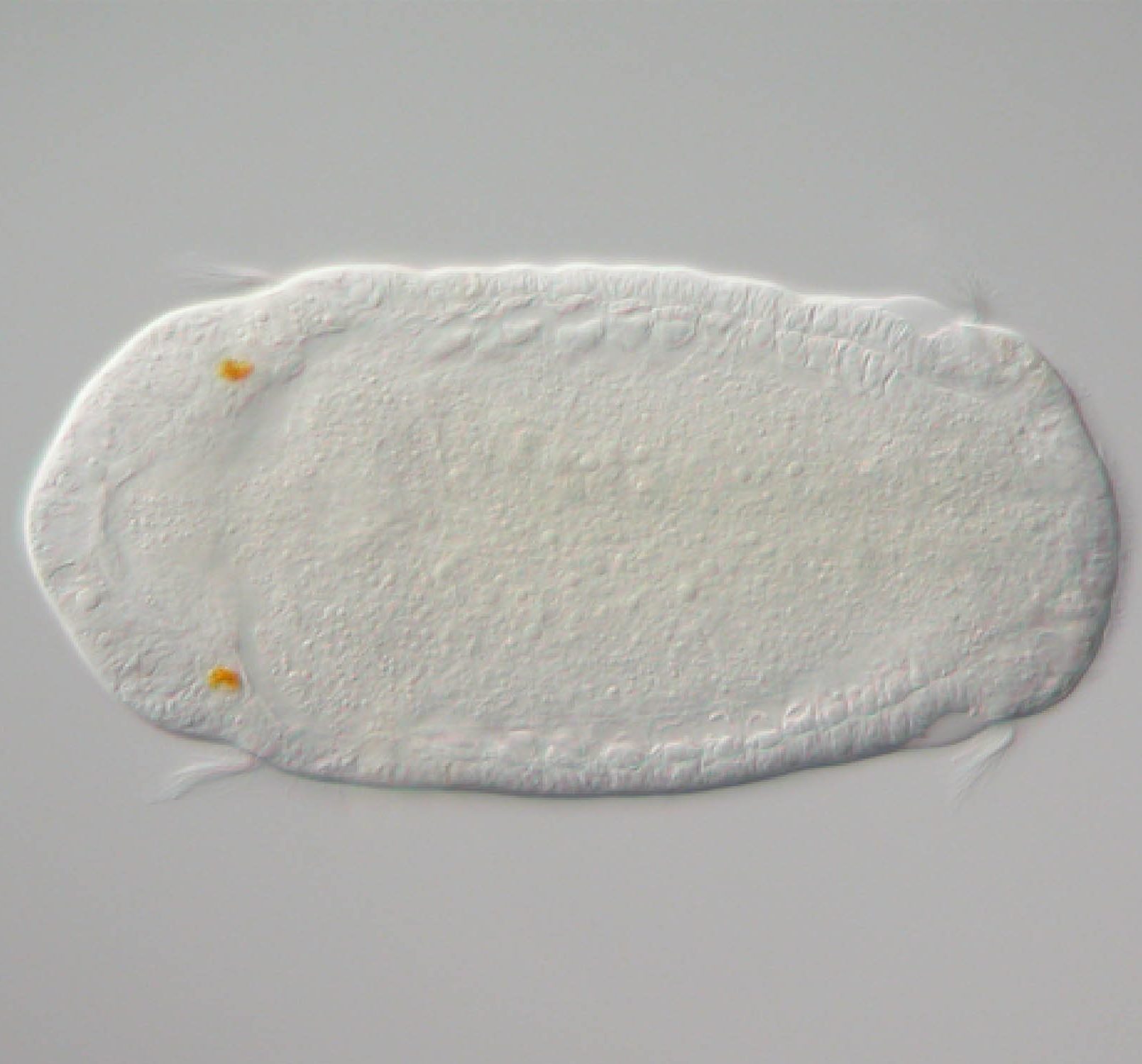
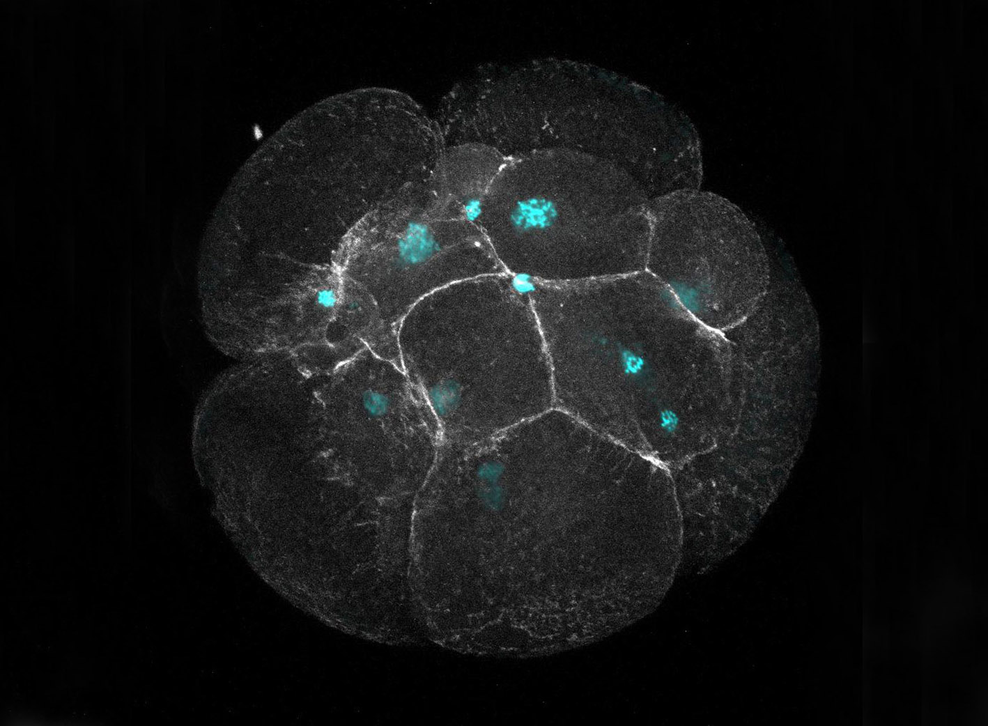
Capitella teleta
Early stage embryo showing identifiable individual cells. Cell outlines are visualized by filamentous actin staining (white) and the nuclei are stained with Hoechst (blue).
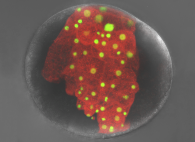
Capitella teleta
Confocal projection of a cleavage stage embryo (22 hr post-injection) in which the C cell was injected at the four cell stage with rhodamine dextran (red) and H2B gfp mRNA (green). At the stage shown, the histone 2B protein has become localized to nuclei.
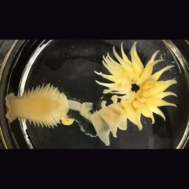
Chaetopterus
Adult with head to the left. This animal has specialized structures along the length of its body that are important for functions such as feeding and reproduction. The animal lives inside a U-shaped tube and filter feeds.
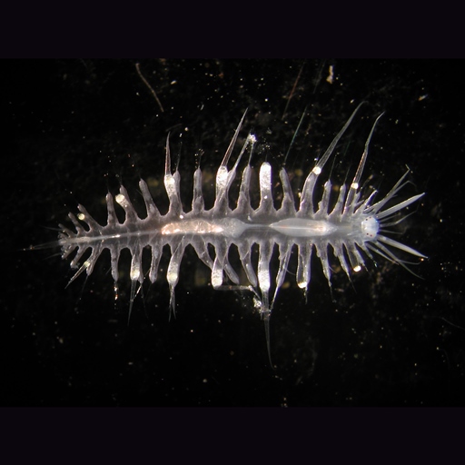
Tomopteris
Adult worm with anterior to the right. Tomopteris is a planktonic annelid and spends its entire life cycle in the water column. Collected off shore of south Oahu.
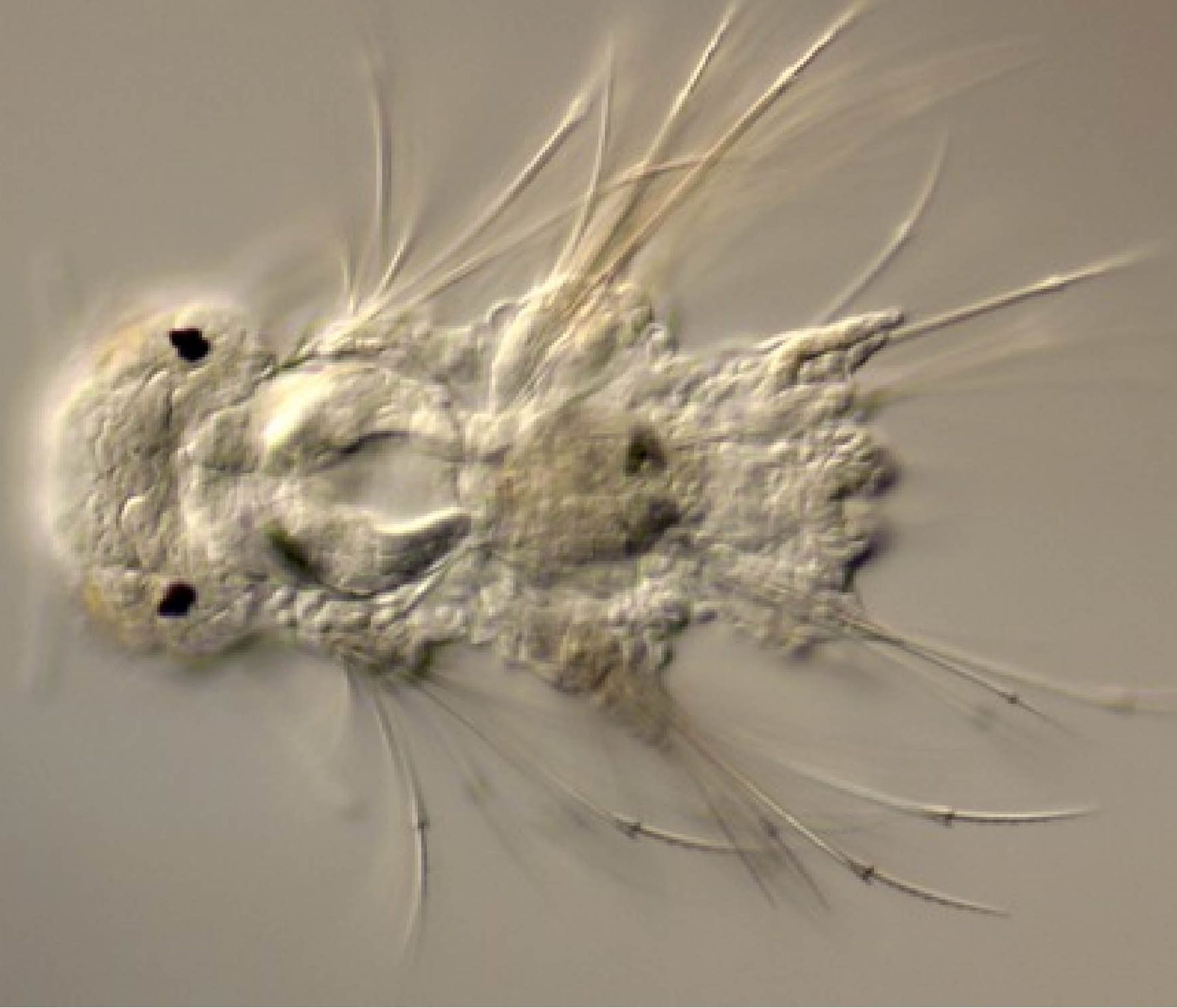
Nereis
Larva with anterior to the left with visible eye spots and chaetae.
Capitella Larvae
Capitella Larvae