Martindale Lab Images
The Martindale Lab works with these fascinating animals.
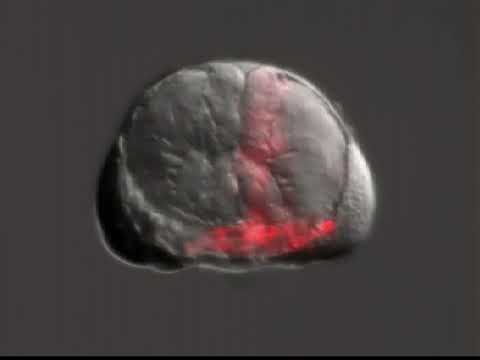
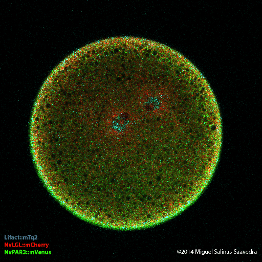
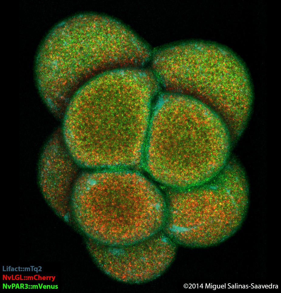
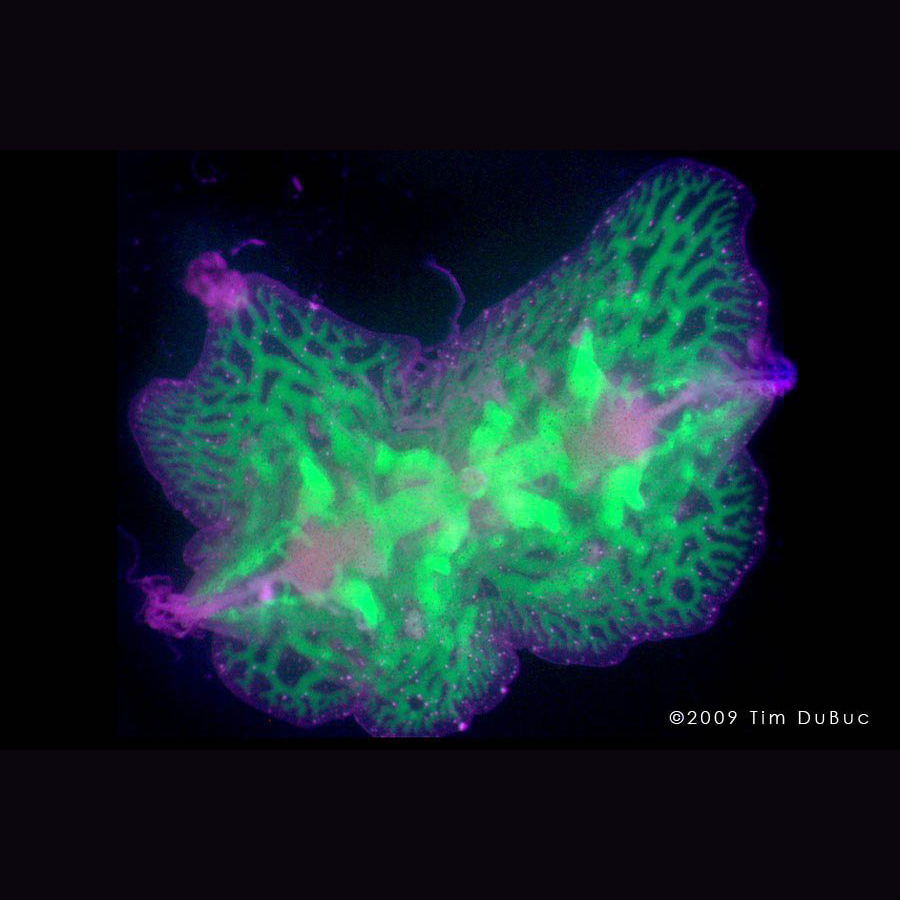
Nematostella Vectensis
PAR-3 and LGL become asymmetrically localized during the early development of Nematostella vectensis.
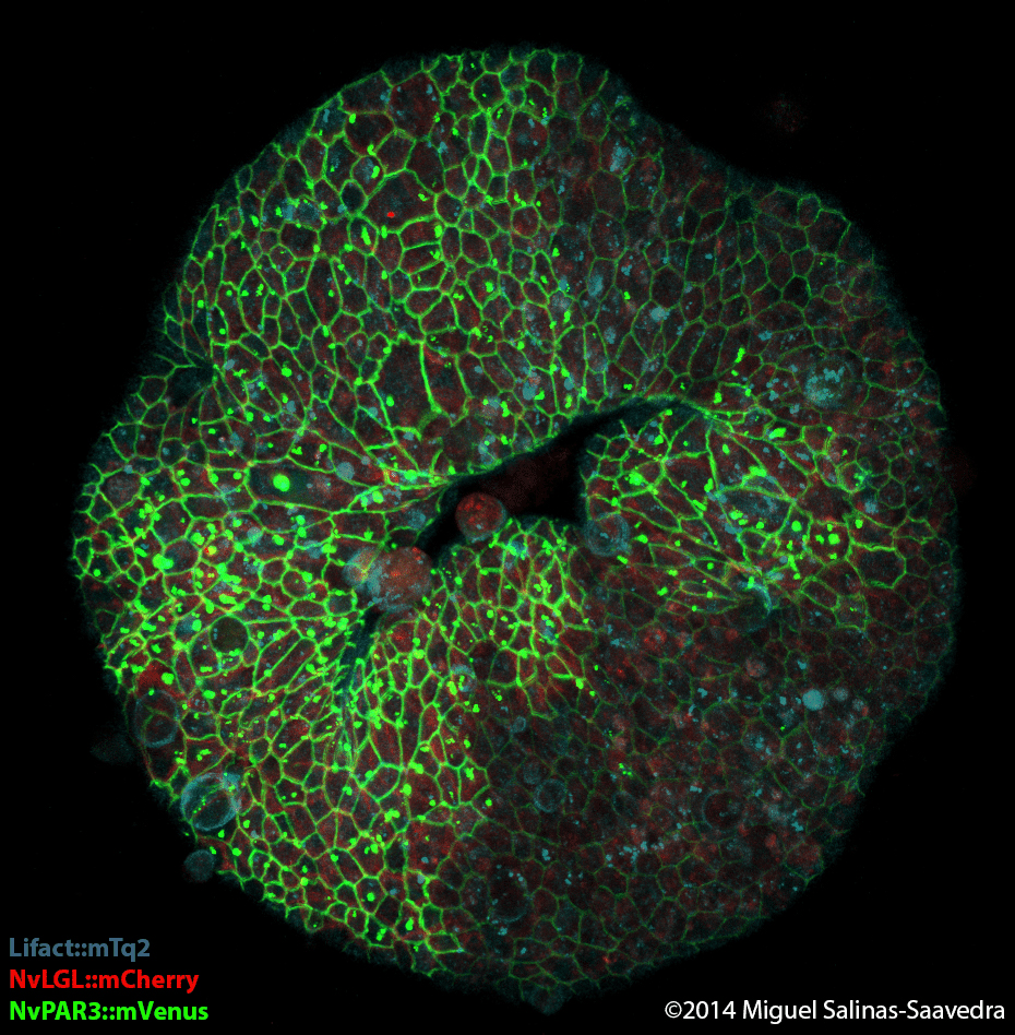
Nematostella Vectensis
PAR-3 and LGL become asymmetrically localized during the early development of Nematostella vectensis.
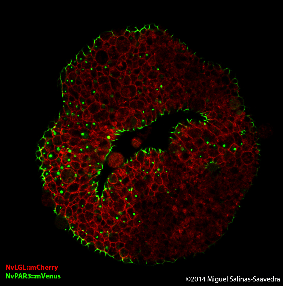
Nematostella Vectensis
PAR-3 and LGL become asymmetrically localized during the early development of Nematostella vectensis.
Nematostella Vectensis
PAR-3 and LGL become asymmetrically localized during the early development of Nematostella vectensis.
Ctenophore
Confocal micrograph of lateral view of a juvenile Florida Beroe ovata ctenophore stained for filamentous actin (green). The apical organ is located at the top of the image and the four tracks of ciliated grooves descend to the tops of the four comb rows seen in this image. Also shown are the complex pattern of muscle cells that run just under the surface of the epidermis. Image taken by David Kainoa Simmons here at the Whitney Marine Lab.
Ctenophore
Confocal micrograph of lateral view of a juvenile Florida Beroe ovata ctenophore stained for filamentous actin (green). The apical organ is located at the top of the image and the four comb rows seen in this image are linked by a complex pattern of muscle cells that run deep under the surface of the epidermis. Image taken by David Kainoa Simmons here at the Whitney Marine Lab.
Ctenophore
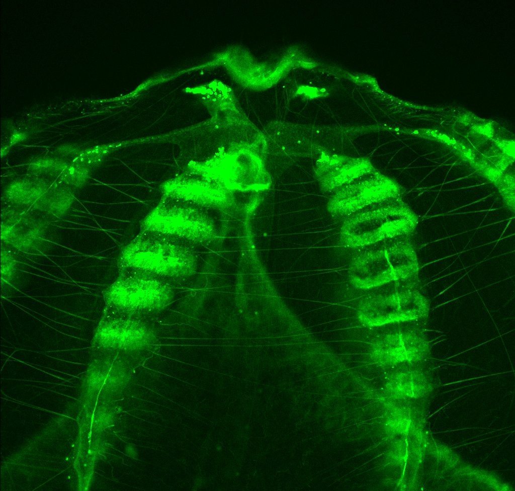
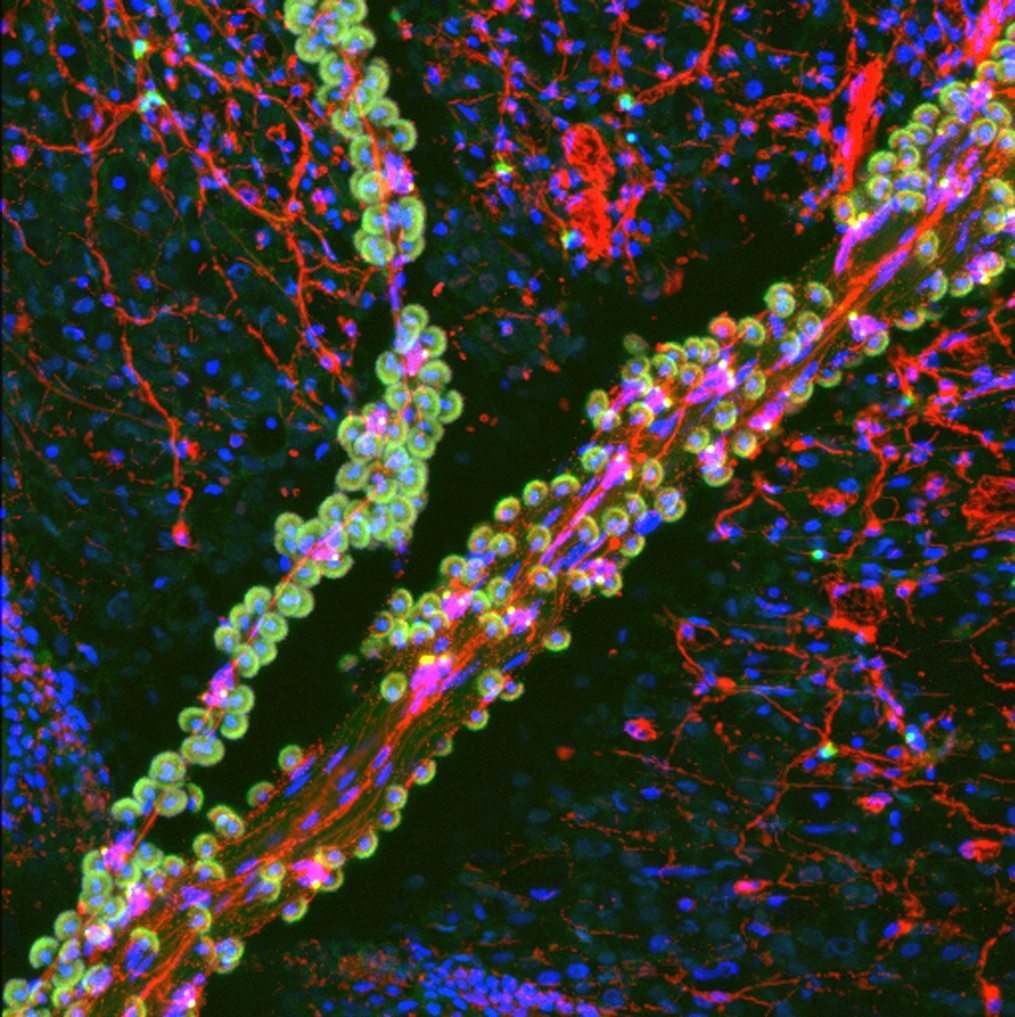
Confocal micrograph of a tentacle of a juvenile Florida Pleurobrachia ctenophore stained for filamentous actin (green) and neurons (red). Nuclei are labeled blue. The tentacle runs from upper right to lower left and contains tentacular nerves and the “sticky” colloblast cells (small green ‘grapes’ with blue nuclei in the center) that attach to prey items. A small branch of the tentacle can also be seen running vertically on the left side of the image. Under the tentacle is the surface of the animal with its chickenwire-like nerve net (red). Image taken by David Kainoa Simmons here at the Whitney Marine Lab.
Ctenophore
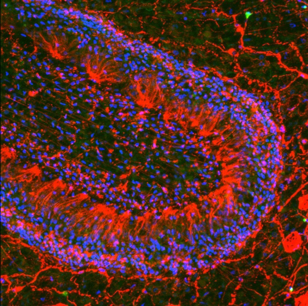
Confocal micrograph of the polar field region of the apical organ of the Florida ctenophore Pleuobrachia. Nuclei are shown in blue and neural elements in red both within, and under the surrounding epidermis (anti tyrosine tubulin). Image taken by David Kainoa Simmons here at the Whitney Marine Lab.
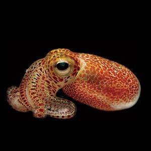
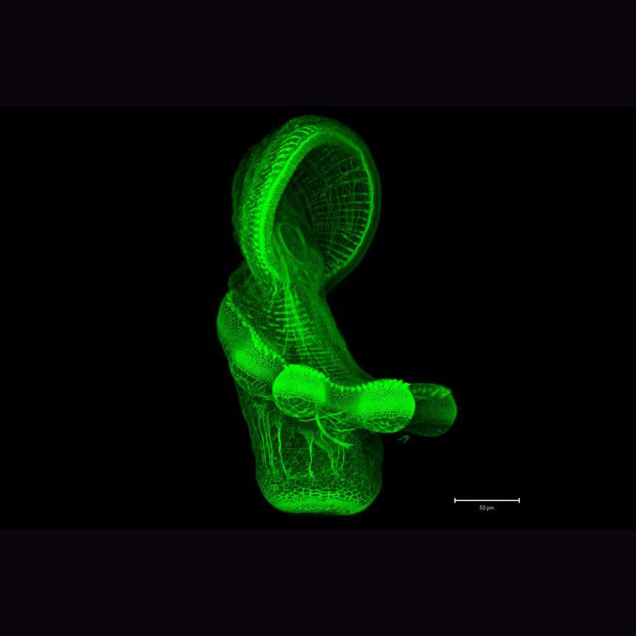
Euprymna Scolopes
The Martindale lab has utilized embryos from a diverse array of species, like this Hawaiian Bobtail Squid (Euprymna scolopes), to understand the evolution of biological novelties.
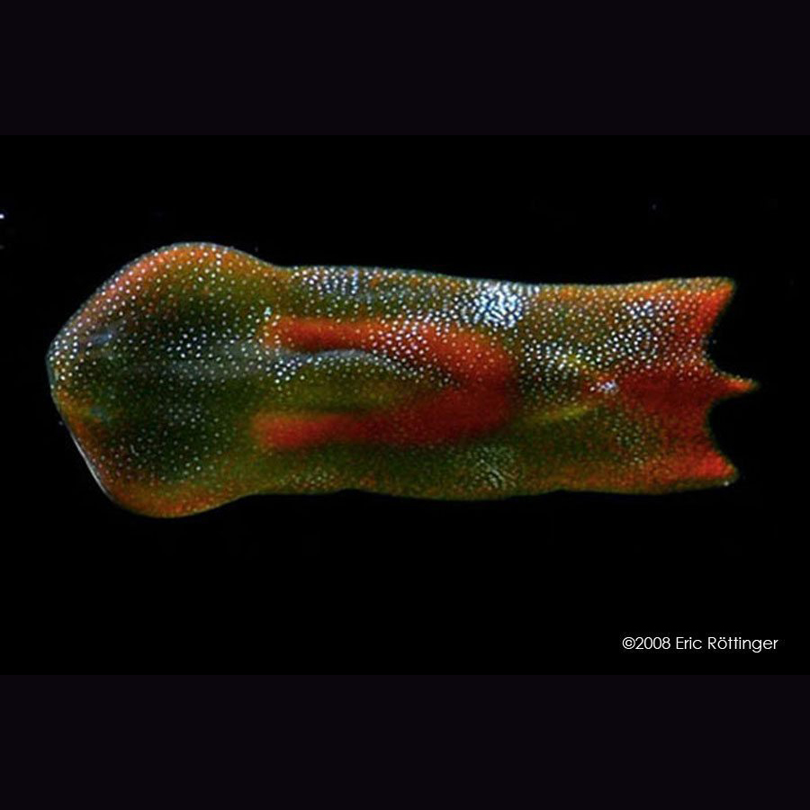
Acoel Flatworm
Embryos of acoelomorphs, like this acoel flatworm, are pivotal for understanding the origin of the anterior-posterior axis, bilateral symmetry, and a definitive mesodermal tissue layer during animal evolution
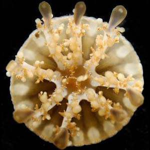
Nematostella Vectensis
Did you know that the genome of this starlet sea anemone, Nematostella vectensis, has more genes in common with human beings than either fruit flies or soil nematodes do (Putnam et al, 2007)?
Benthic Ctenophore
This benthic ctenophore (Coeloplana sp) crawls around on the ocean floor and is an example of a group of animals that are likely to be the earliest branching extant clade of animals.
Cassiopea
“Upside Down Jellyfish” (called Cassiopea) is being used as a new model for symbiosis and coral bleaching.
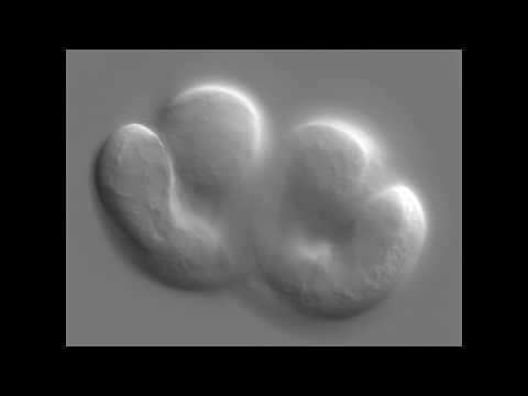
Nematostella Vectensis
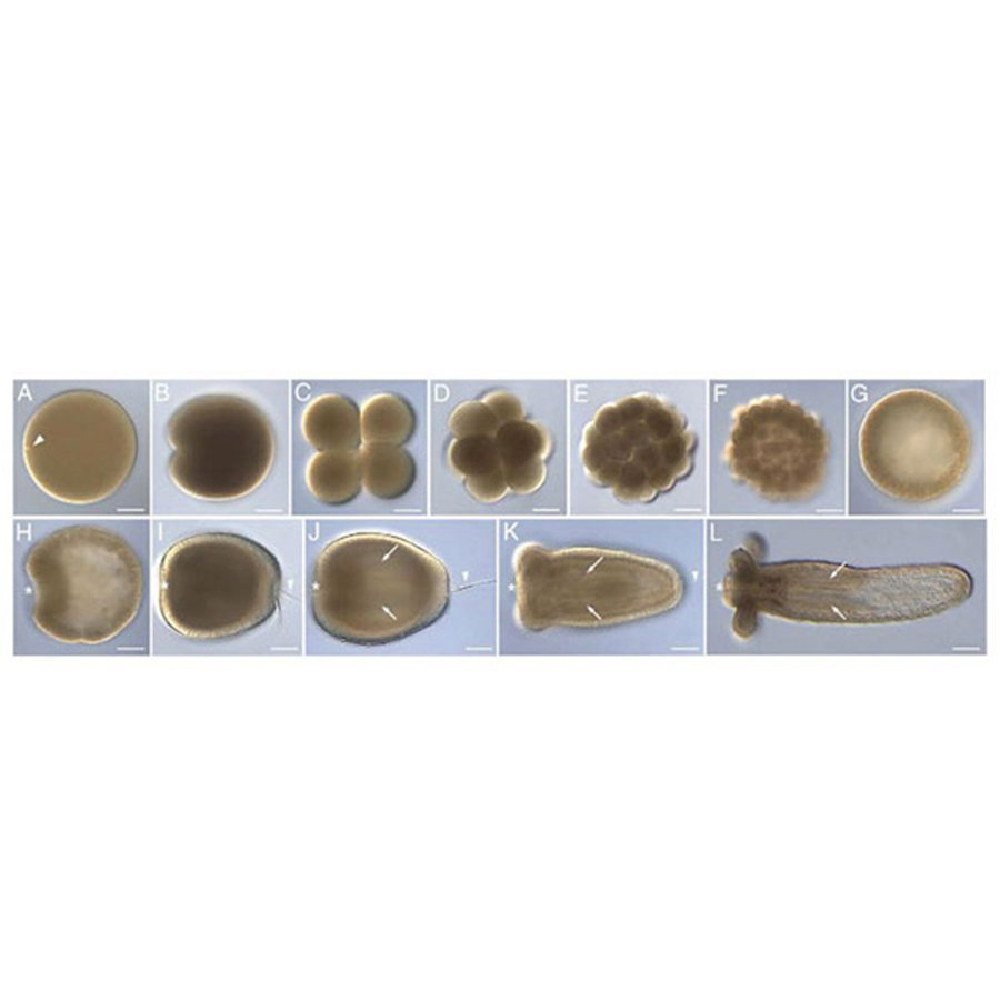
These micrographs of different stages of the development of the sea anemone Nematostella vectensis show that their embryos look very similar to other animal embryos.
Phoronid
This is a confocal micrograph of the actinotroch larval stage of a phoronid whose muscle cells have been visualized with an actin binding dye. The oral hood (top) has opened up to reveal the larval mouth.
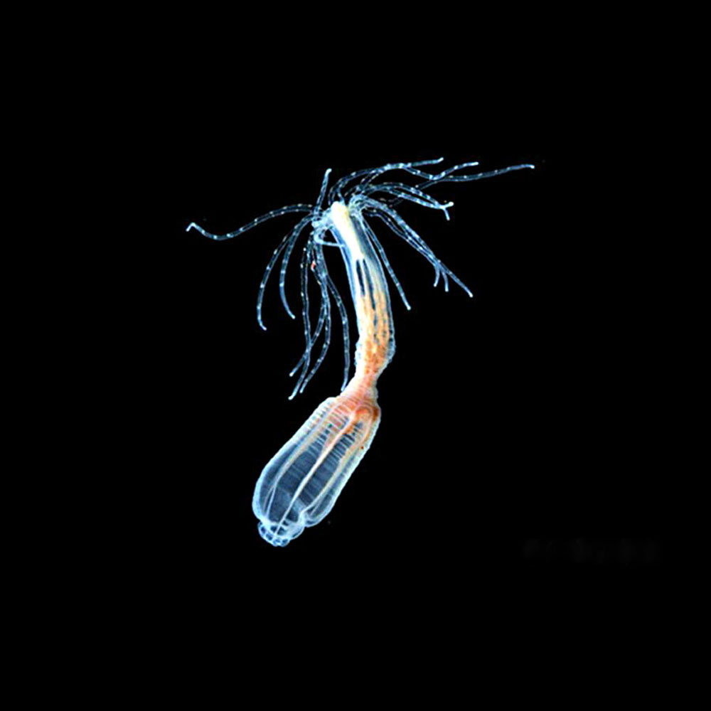
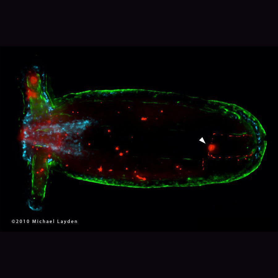
Nematostella Vectensis
This is an image of a live transgenic sea anemone (Nematostella vectensis) generated by former Whitney postdoc Dr. Michael Layden showing a subset of its nervous system expressing the green fluorescent protein (GFP). The mouth and oral tentacles are seen to the left.
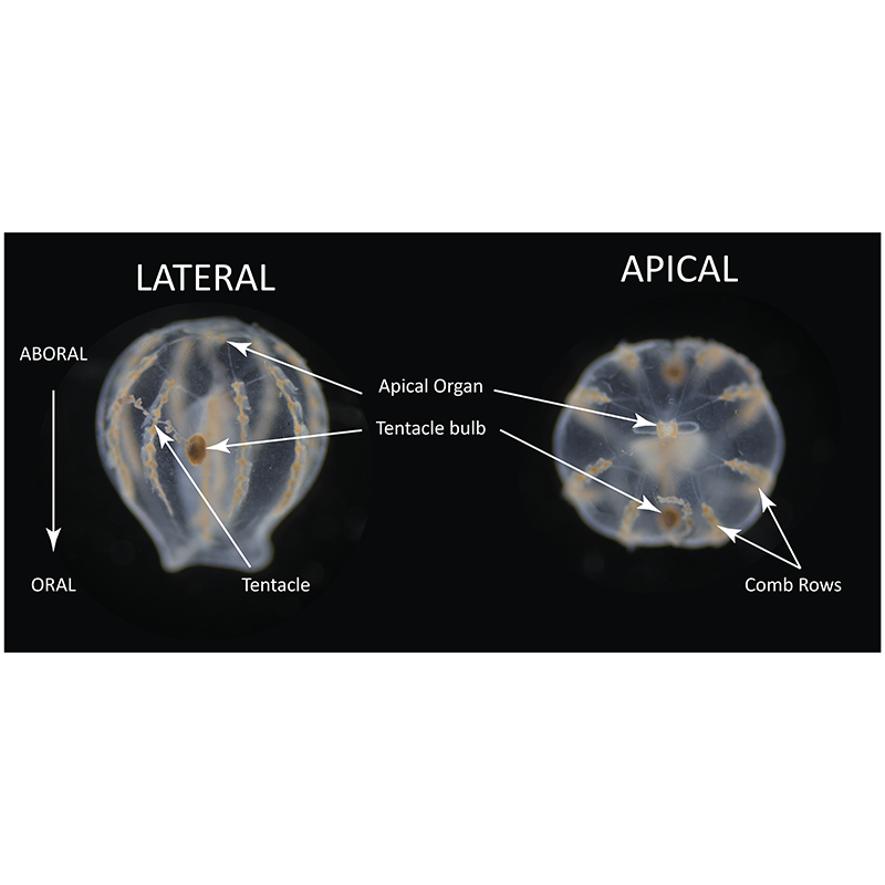
Mnemiopsis Leidyi
Gross morphology of the cydippid stage ctenophore Mnemiopsis leidyi. Animals were chemically fixed prior to imaging. Picture by Dorothy Mitchell
Mnemiopsis Leidyi Cydippid
Immunofluorescent staining of a whole Mnemiopsis leidyi cydippid. Animal is in the apical position and features DAPI(blue), anti-actin(green), and anti-tyrosinated tubulin. Photo by Dorothy Mitchell