UF Whitney Laboratory biologists show how a segmented marine worm found locally in muddy estuaries gradually gains the ability to regenerate during its life cycle. Their results highlight how developmental timing impacts the ability to regenerate missing tissues and structures, introducing a new angle on how regenerative abilities can change across time and hinting at potential mechanisms that could in theory be useful for switching regeneration on and off.
Regeneration generally requires three basic steps: first, close off the wound; second, recruit cells that have the potential to recreate the missing tissue; and third, regrow the lost structure. How those steps are implemented can vary substantially between species.
The segmented worm, Capitella teleta, can regenerate its tail but not its head as an adult. PhD candidate Alicia Boyd decided to see if larvae had the same regenerative abilities. As part of her inspiration, Boyd points to frogs that can regenerate their tails as tadpoles and then lose that ability with age.
Using very thin glass needles, she cut off the heads of some of her larvae and the tails of others. She noticed that dividing cells localize at both cut sites within six hours. After two days, a stem cell marker gene called vasa shows up at the cut sites, potentially to begin the process of recreating missing tissue. However, cells in head regions seemed to get stuck as they tried to transform into neurons — they weren’t quite able to complete the transition.
In the end, larvae could regrow tissue in their tails, but not their heads — similar to adults. However, larvae weren’t able to fully regrow every missing structure. Taken together, these observations indicate that larvae can begin early stages of regeneration but not finish it. This suggests C. teleta gradually increases in regenerative ability throughout its lifecycle, opposite to what’s seen in frogs.
These results were reported in an online Early View of Invertebrate Biology on Oct. 20.
Investigating the developmental onset of regenerative potential in the annelid Capitella teleta
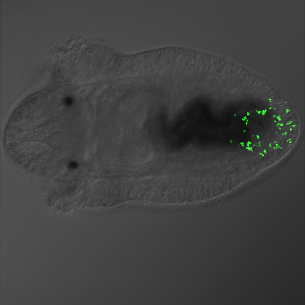
Image: An amputated Capitella teleta larva, five days after amputating the tail. Dividing cells (green) are grouped at the cut site, indicating birth of new cells. In animals capable of regeneration, birth of new cells represents an early stage of the regeneration process. The larva is approximately 325 um. (photo credit: Alicia Boyd)