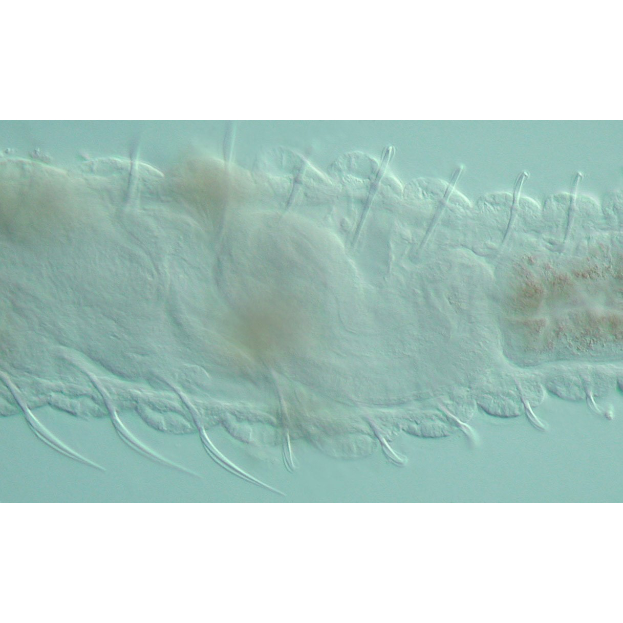
Segmented worms are commonly found in our local marine environment, both in estuaries and in the open ocean. One of the defining features of the 20,000+ described species of segmented worms is the presence of chitinous bristles that stick out from the body wall and often aid in locomotion and anti-predatory behavior.
A new paper from the Seaver Lab characterizes chaetal formation in the segmented worm Capitella teleta. Most published studies of chaetal formation focus on adult animals. This morphological study is unique in that it focuses on the earliest formation of chaetae earlier in the life cycle, specifically in larvae and young juvenile worms. The surprising finding from this study was the identification of a transient glandular structure associated with the chaetae in larvae but not in adult animals. This newly identified structure has not been described in any other annelid to date and its exact function remains somewhat of a mystery.
The manuscript entitled ‘Discovery and characterization of a transient chaetal gland during the development of Capitella teleta (Sedentaria: Annelida)’ is an international collaboration with Dr. Ekin Tilic and Dr. Thomas Bartolomaeus, both world experts on chaetal formation. The study was published on June 4 in the Journal of Morphology.
Image: Midbody region of a young juvenile worm of the annelid Capitella teleta (3 days post-metamorphosis). Chaetae project from each side of the body. In this image, pairs of chaetae are visible in each body segment. The length of the region shown is approximately 700 microns and the anterior end of the worm is to the left.