Congratulations to Justin Waletich, Dr. Gonzalo Quiroga-Artigas (previous Schnitzler Lab postdoc), Dr. Danielle de Jong, and Dr. Christine Schnitzler who published a paper with collaborators titled “The genome of the colonial hydroid Hydractinia reveals their stem cells utilize a toolkit of evolutionarily shared genes with all animals” in the journal Genome Research. This paper represents the result of a large-scale international collaborative effort to sequence the genomes of two species of the model hydroid, Hydractinia and perform comparative analysis to place these genomes in a comparative evolutionary framework with other cnidarians and other animals. A companion website, the Hydractinia Genome Project portal, https://research.nhgri.nih.gov/hydractinia/ provides access to a comprehensive set of resources for exploring the genome and related data from the publication.
Hydractinia is a colonial marine hydroid that exhibits remarkable biological properties, including the capacity to regenerate its entire body throughout its lifetime, a process made possible by its adult migratory stem cells, known as i-cells. Here, we provide an in-depth characterization of the genomic structure and gene content of two Hydractinia species, H. symbiolongicarpus and H. echinata, placing them in a comparative evolutionary framework with other cnidarian genomes. We also generated and annotated a single-cell transcriptomic atlas for adult male H. symbiolongicarpus and identified cell type markers for all major cell types, including key i-cell markers. Orthology analyses based on the markers revealed that Hydractinia’s i-cells are highly enriched in genes that are widely shared amongst animals, a striking finding given that Hydractinia has a higher proportion of phylum-specific genes than any of the other 41 animals in our orthology analysis. These results indicate that Hydractinia’s stem cells and early progenitor cells may use a toolkit shared with all animals, making it a promising model organism for future exploration of stem cell biology and regenerative medicine. The genomic and transcriptomic resources for Hydractinia presented here will enable further studies of their regenerative capacity, colonial morphology, and ability to distinguish self from non-self.
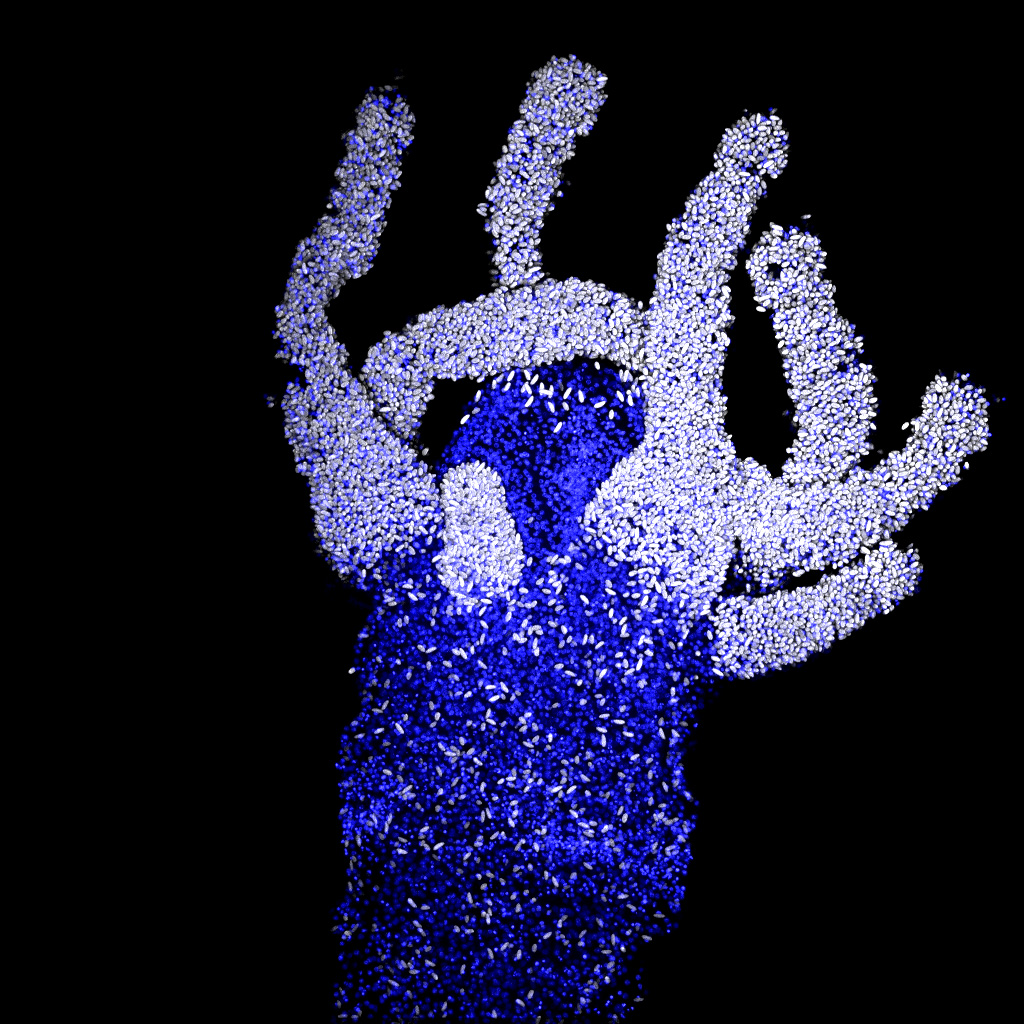
Image: Hydractinia symbiolongicarpus adult feeding polyp detached from the colony. The white cells are cnidocytes (stinging cells) highlighted using High DAPI staining. Blue cells are stained with POPO1, a nuclear dye. Image by Gonzalo Quiroga Artigas.